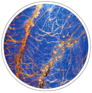
Qubit Systems offers non-invasive root screening of plants from early seedling emergence to late in plant development. Plants are grown in Rhizotubes with roots visible when outer light shield is removed. The whole system is delivered to the RhizoCab and rotated during imaging. High resolution images allow visualization and morphometric analysis of the whole root system resulting in detailed root phenotyping opportunities. Combined with the PlantScreen systems whole plant phenotyping is possible.
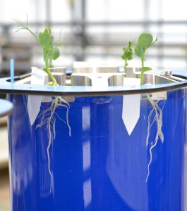
Image the Entire Root System for Non-Invasive Assessments of:
- Biotic and Abiotic Stress
- Nutrient Regimes
- Soil Structure
- Plant – Microbe Interactions etc.
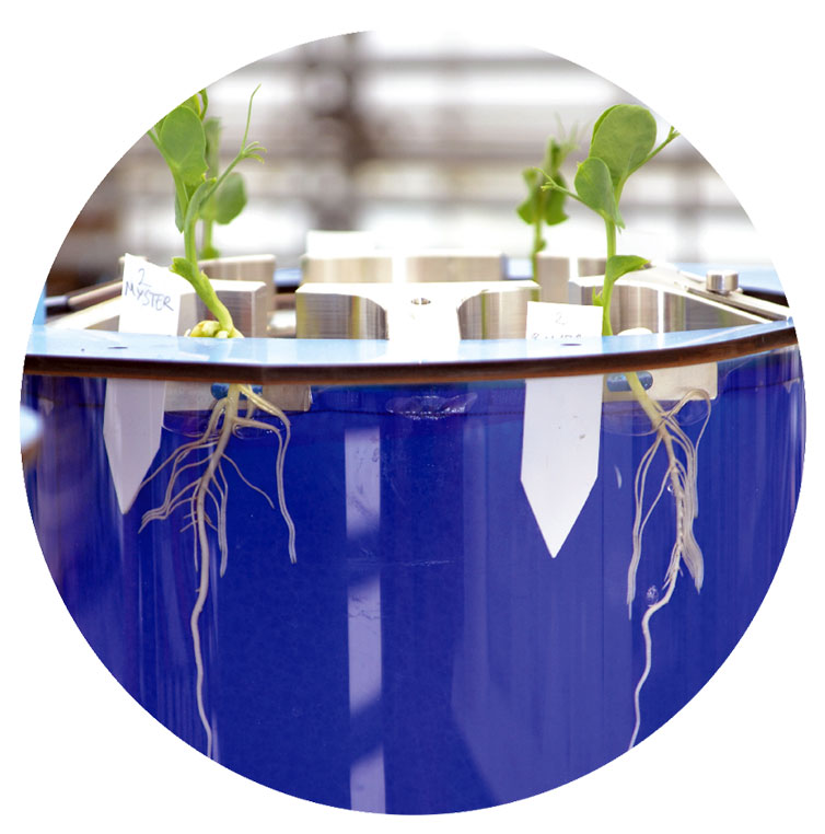
Roots are visible and measurable immediately after emergence.
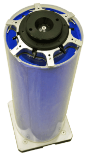
The Rhizotube is a cylindrical rhizotron that consists of two concentric tubes. Plants are grown between the outer tube and a plastic membrane, 18 um thick, that allows passage of water, nutrients and microorganisms to the root, but does not allow the root to penetrate to the growth substrate between the mesh and the interior tube. Therefore, the entire root system is visible for imaging. A light shield surrounds the rhizotube during plant growth to prevent the establishment of algae.
One to six plants may be grown in a single Rhizotube. The growth substrate is added using a machine that ensures homogenous compaction and minimises variablity between Rhizotubes. Nutrient solution is added via an automated nozzle and is distributed evenly to the plants via a manifold system. An automated drainage valve ensures that the substrate never becomes waterlogged (unless the researcher wishes to study flooding).
Rhizotubes may be delivered by conveyor, or loaded automatically, into the Rhizocab imaging cabinet. The Rhizotube is rotated to obtain a complete scan of the root system using a side mounted 6 Megapixel camera. The Rhizotube is illuminated in sequence with red, blue and green light via an LED light source, to provide ultra high definition colour images. The camera may be moved automatically on a linear motor to zoom in on a specific area of the root to provide images at even higher definition. The resolution of images ranges from 42 μm per pixel at 600 ppi to 7 μm per pixel at 3600 ppi. This allows visualisation and morphometric analysis of the thinnest roots, as well as detection of nodules and mycorrhizae.
Each Rhizotube stands 50 cm high and can accommodate both large and small plants. A heavy metal base ensures a low centre of gravity so that the Rhizotrons may be transported on a conveyor system to the imaging cabinet. Rhizotron design allows for both root imaging and optional shoot imaging with Qubit’s PlantScreen Phenotyping System.
Rhizotubes and the Rhizocab (patents pending) were developed at UMR Agroécologie (INRA Dijon, France) in collaborations with InoviaFlow (Dole, France). Qubit Systems Inc. distributes the Rhizotube and Rhizocab technologies worldwide.

Extensive validation of the Rhizotube method for root screening has been undertaken by Dr. Christophe Salon and colleagues at INRA UMR Agroecologie in Dijon, France. The work was published in 2016: RhizoTubes as a new tool for high throughput imaging of plant root development and architecture: test, comparison with pot grown plants and validation. Jeudy, C. et al. 2016 Plant Methods.
